Medit Model Builder is an innovative and powerful tool for creating physical models from oral scanner data. With its advanced ability to transform digital information into highly accurate physical models, Medit Model Builder offers an efficient and practical solution for professionals seeking quality and accuracy in their dental models.

Creating Physical Models with Oral Scanner Data
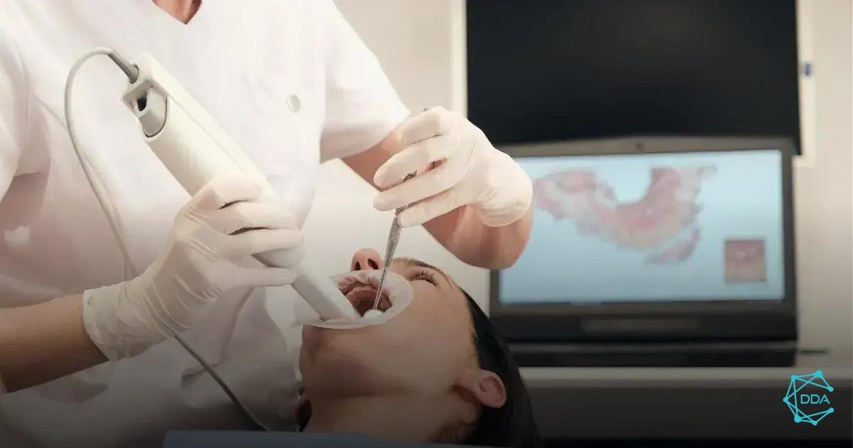
Creating physical models from oral scanner data is an essential process in modern dentistry. These models provide an accurate representation of a patient's oral structure, allowing dental professionals to visualize anatomical details and plan procedures with greater precision.
To create physical models from oral scanner data, you need to follow a set of careful and precise steps. The quality and accuracy of the resulting model depends on attention to detail at each stage of the process.
Oral Scanner Data Collection
The first step in creating a physical model is collecting data from the oral scanner. This process involves using advanced technology to capture a three-dimensional digital representation of the patient's oral structure.
The data collected by the oral scanner provides detailed information about the geometry and topography of the oral region, including the surrounding teeth, gums, and bony structures.
3D Processing and Modeling
After collecting data from the oral scanner, the next step is processing and 3D modeling. At this stage, the captured data is processed by specialized software to create a three-dimensional digital model of the patient's oral structure.
This digital model serves as the basis for creating the physical model, and it is essential to ensure accuracy and fidelity to the original data during the modeling process.
3D Printing of the Physical Model
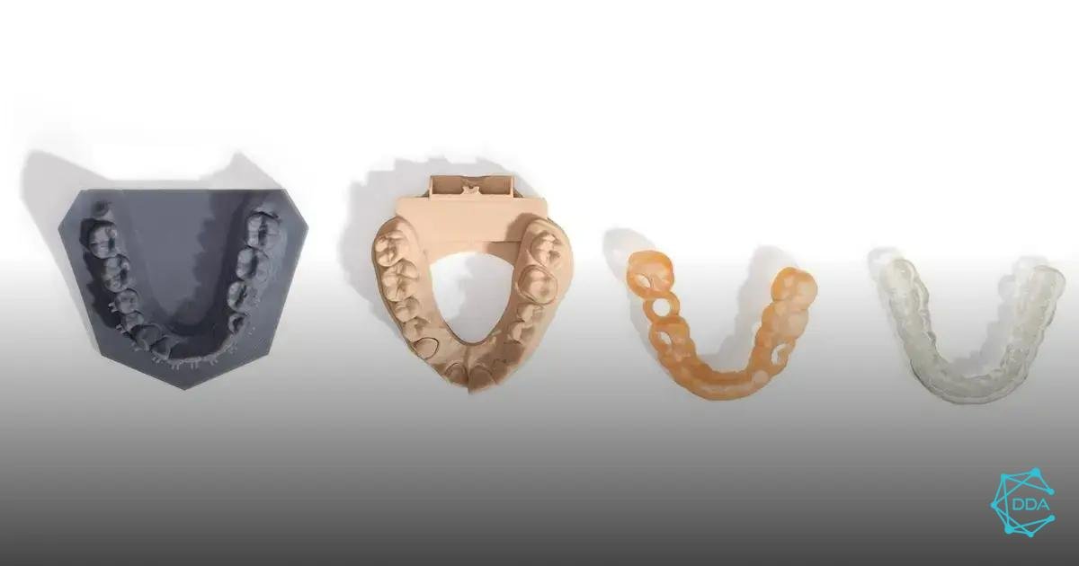
With the 3D digital model finalized, the next step is 3D printing of the physical model. This process involves using 3D printing technology to transform the digital model into a solid physical model.
The choice of impression material is crucial to ensuring the accuracy and durability of the physical model, and should be selected based on the specific needs of the planned dental procedure.
In summary, creating physical models with oral scanner data is a complex process that requires attention to detail and the use of advanced technology. When carried out accurately, this process offers dental professionals a valuable tool for planning and executing procedures with greater precision and effectiveness.

Attaching Articulator for Verification
The step of attaching the articulator for verification is crucial in the process of creating physical models from oral scanner data. It is essential to ensure that the articulator is correctly positioned so that the modeling is accurate and faithful to the patient's anatomical reality.
To carry out this step successfully, it is important to follow the following steps:
- Articulator Preparation: Before attaching the articulator, it is essential to ensure that it is clean and in perfect working order. Any misalignment or dirt can compromise the accuracy of the final model.
- Attachment to the Oral Scanner: The articulator must be properly fixed to the oral scanner, ensuring that it remains stable throughout the scanning process. This avoids distortions in the captured data.
- Position Check: After scanning, it is necessary to check whether the articulator is correctly positioned and aligned with the scanner data. Any misalignment must be corrected before proceeding to the next step.
Once the articulator has been correctly attached and verified, the modeling process can proceed with greater confidence in the accuracy of the data obtained.

Labeling Bases for Model Distinction
The step of labeling the bases for distinguishing models is crucial in the process of creating physical models from oral scanner data. The correct identification and marking of the bases guarantees the precision and quality of the final model.
It is important to highlight that labeling the bases allows differentiation between models and correct assembly in the articulator to verify occlusion.
To carry out labeling, it is necessary to follow a marking standard that allows clear identification of the parts. This includes the use of codes or symbols that represent the specific characteristics of each base, such as the dental arch, the anterior and posterior regions, among others.
Furthermore, the use of different colors for the different bases can facilitate visual identification and speed up the assembly and verification process. Correct labeling of bases significantly contributes to the efficiency and accuracy of the physical model creation process.


Practical Demonstration of Use
The practical demonstration of the use of physical models with oral scanner data is an essential step to understanding the process of creating and using these models. During the demonstration, professionals have the opportunity to view in detail how models are created from data obtained from the oral scanner.
It is during this stage that professionals can verify the accuracy and quality of physical models, as well as understand how data from the oral scanner is converted into three-dimensional models.
Furthermore, the practical demonstration of use also allows professionals to learn how to handle and use the articulator to check occlusion on physical models. This is essential to ensure the accuracy and functionality of the models in clinical practice.
Another important aspect of the practical demonstration is the labeling of the model bases, which allows distinction between different models and facilitates the process of identification and use in the day-to-day practice.
In summary, the practical demonstration of use provides professionals with the opportunity to experience first-hand the entire process of creating, verifying and using physical models with oral scanner data, preparing them to apply this knowledge in their professional practice.

